Questions
- What is X-Ray Crystallography?
- ==X-ray crystallography is a technique used to determine the three-dimensional structure of molecules, particularly large biomolecules such as proteins and nucleic acids==.
The technique involves the use of X-rays, which are electromagnetic radiation with wavelengths shorter than visible light, to create a diffraction pattern of the molecule. - The process of X-ray crystallography typically involves several steps:
- Purification of the molecule of interest, such as a protein or nucleic acid.
- Crystallization of the molecule, which involves the formation of a crystal lattice of identical molecules.
- Collection of X-ray diffraction data, which involves exposing the crystal to a beam of X-rays and measuring the resulting diffraction pattern.
- Analysis of the diffraction data using computational methods to determine the electron density map of the molecule.
- Model building, refinement, and validation of the three-dimensional structure of the molecule.
- The resulting three-dimensional structure of the molecule can provide valuable insights into its function, as well as guide the development of new drugs and other therapeutics.
- X-ray crystallography has been used to determine the structures of many important biological molecules, including the DNA double helix, the ribosome, and numerous proteins involved in diseases such as cancer and Alzheimer’s.
It is a powerful tool in the field of structural biology and continues to play a critical role in drug discovery and other research fields.
- ==X-ray crystallography is a technique used to determine the three-dimensional structure of molecules, particularly large biomolecules such as proteins and nucleic acids==.
—————————————————————
IMPORTANTE
IMPORTANTE X-ray Crystallography:
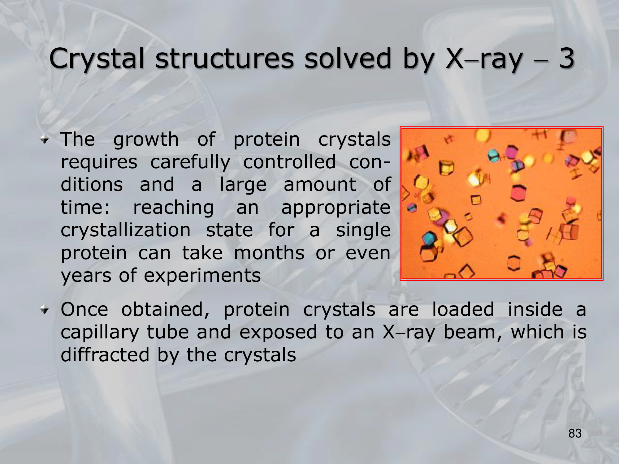
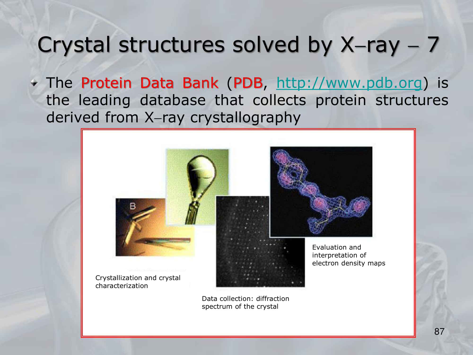
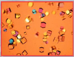
- Crystallize your protein. It takes a lot of time (months or years) and it’s a really delicate process, crystals are grown by the evaporation of a solution of pure protein (like salt and sugar cristals), the final product is more of a gel than a crystal, it’s very small ~ mm (in each direction), also the crystals are composed of about water.
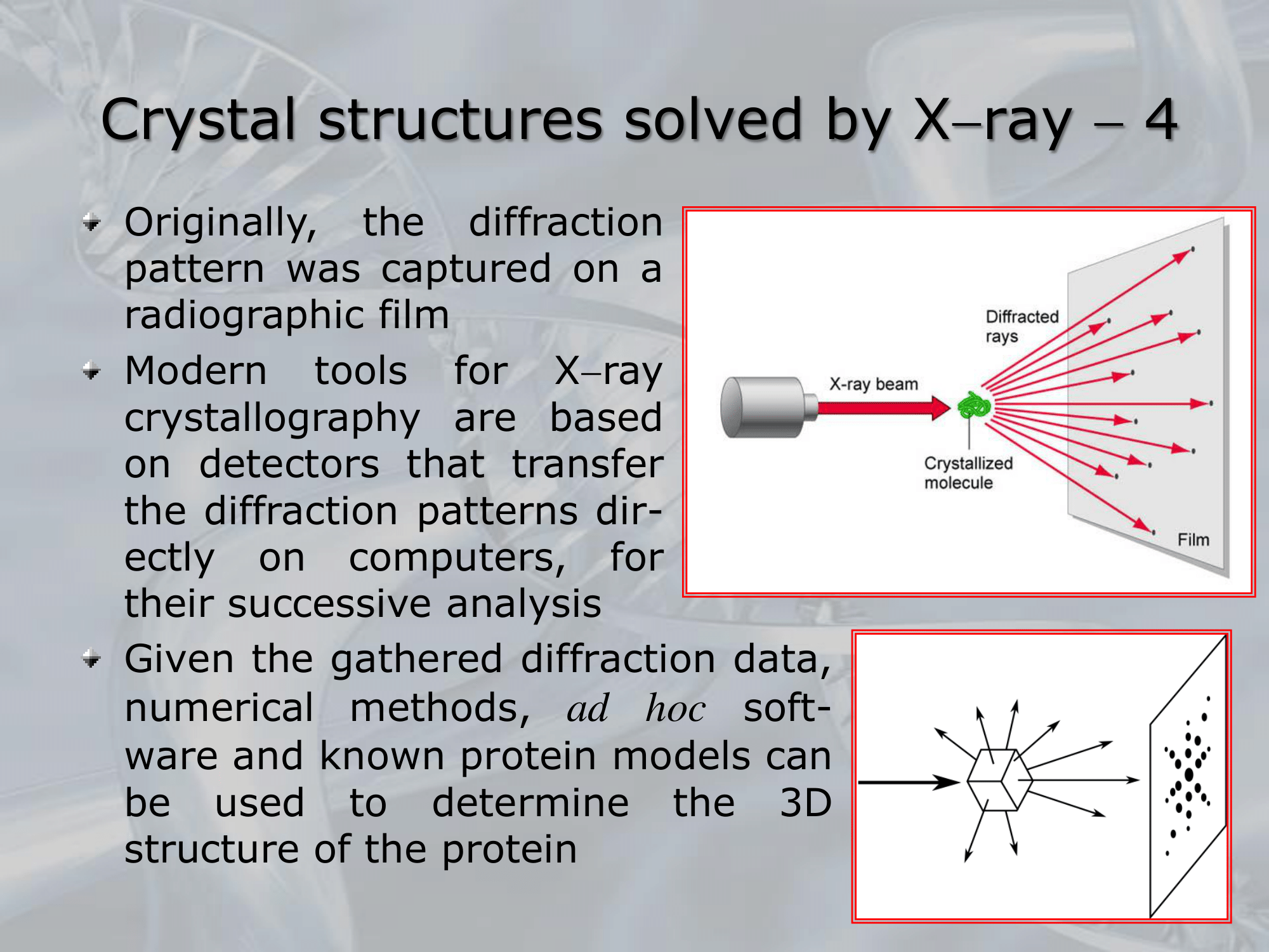
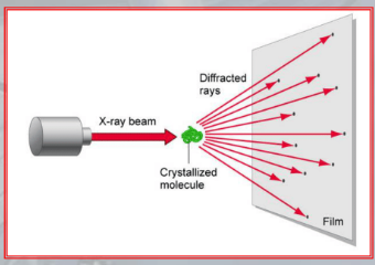
- The crystal are loaded insed a capillary tube and exposed to an X-ray beam, which is diffracted by the crystals.
- The beam is than captured on a radiographic film (today we use detectors to transmit data directly to computers).
- Using ad hoc algorithms, we take the point of the diffracted rays and we obtain a highly accurate 3D model of the protein.
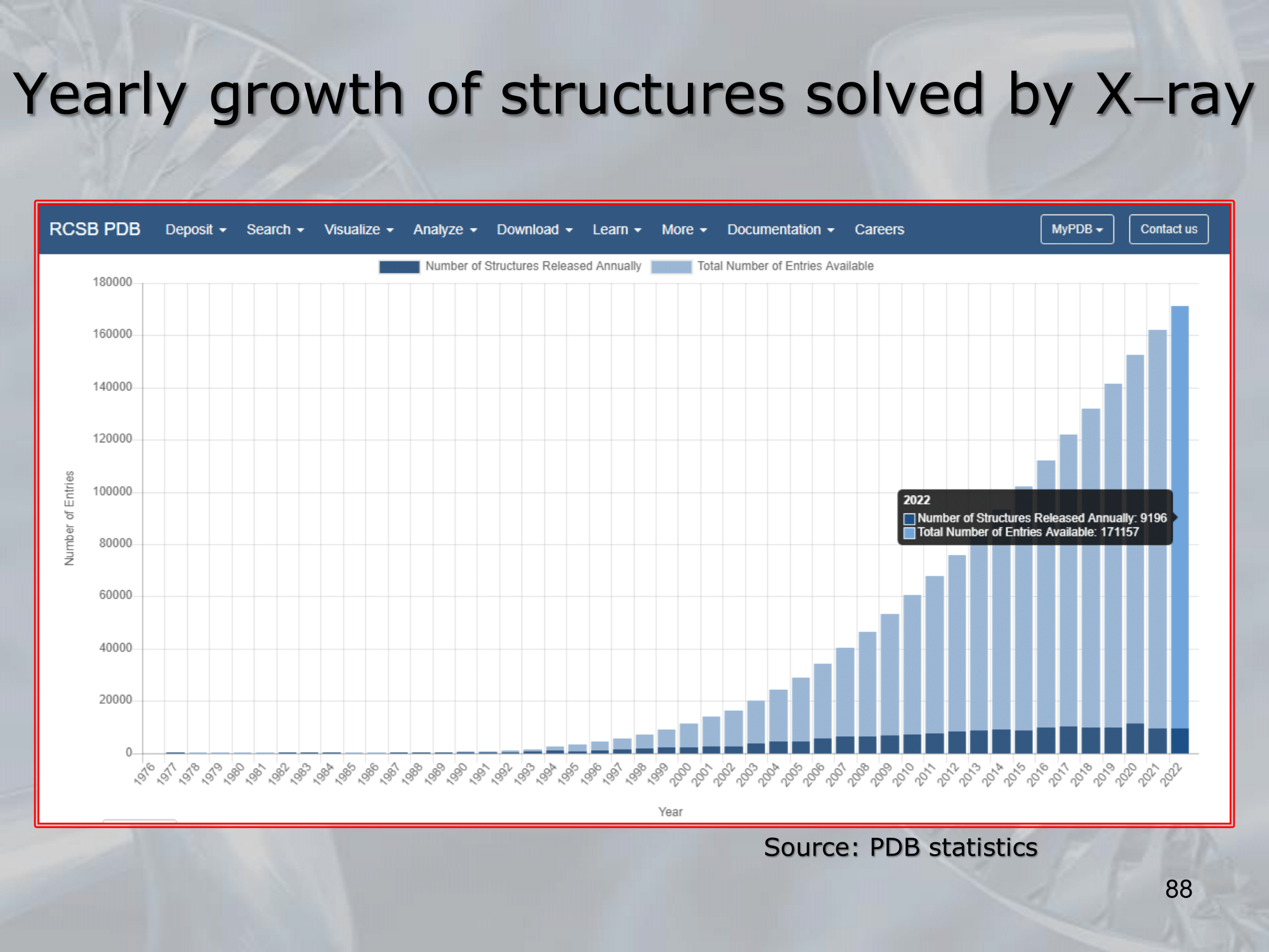
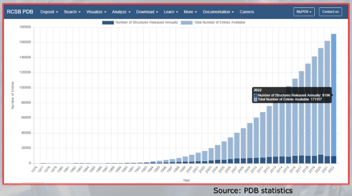
In detail from the X-ray diffreaction spectrum of the crystal, we are able to calculate the electron density map, which is in practice an upscaled image of the molecules from the crystal (upscaled ) Then a computer we verify the 3D structure agreement (fitting) with a molecular model. The error is between ~ (really good) The final structure is the avarage of multiple crystallographies of copies of a single protein, the protein are not compleatelly rigid, and we need to eliminate the background noise produced by the water molecule in each crystal, so this step is necessary. Crystallography is still the main way to get 3D protein Structures, as of now proteins have been analyzed by x-ray crystallography.
—————————————————————
Slides with Notes