Questions
- What are Electron Microscopy Structures?
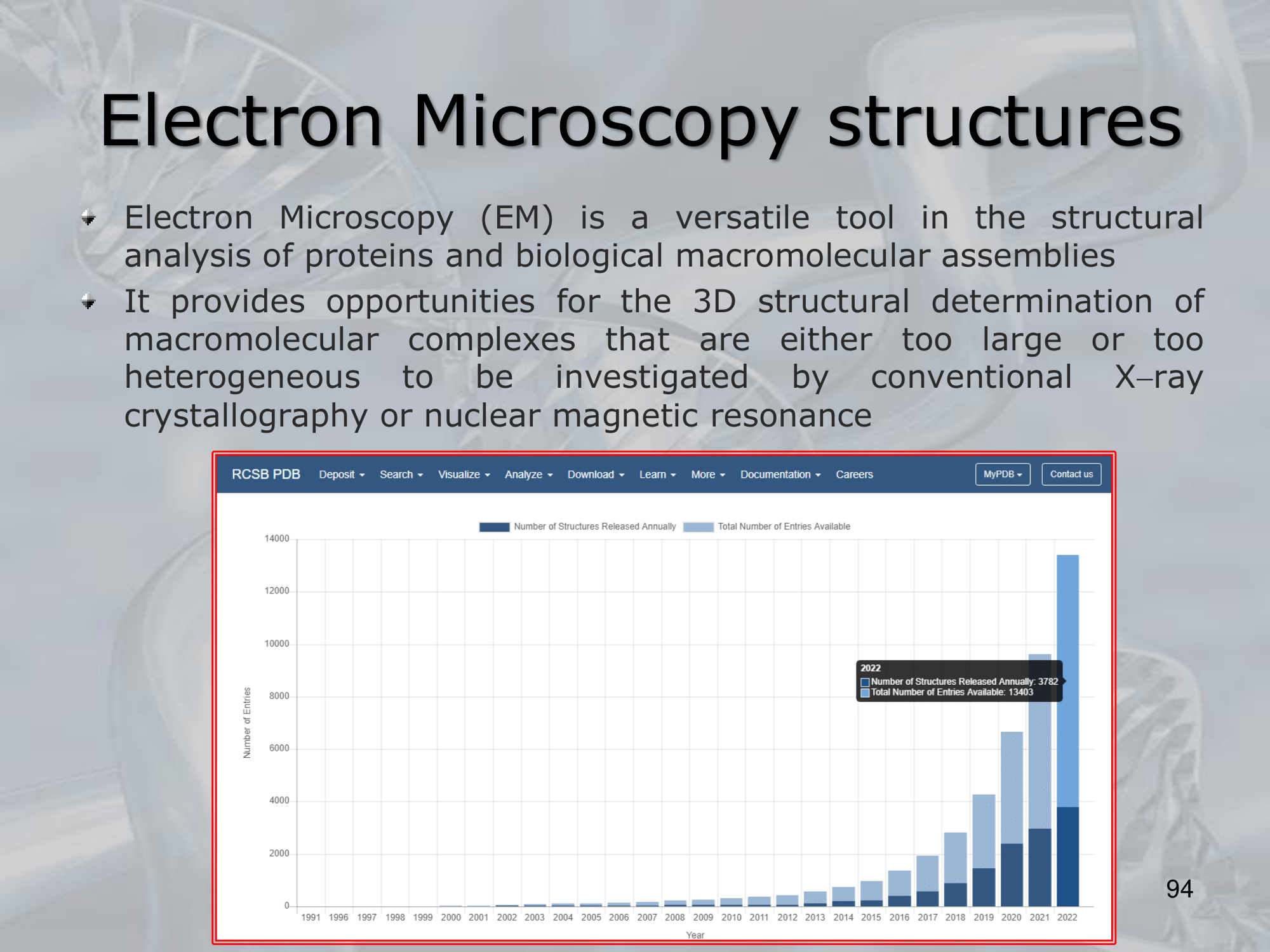
- ==Electron microscopy (EM) is a technique used to visualize the structures of biological molecules and complexes at high resolution==.
In the context of bioinformatics, EM structures refer to structures of these biological molecules that have been determined using electron microscopy. - EM works by passing a beam of electrons through a sample of the biological molecule or complex, which scatters the electrons and produces a pattern that can be recorded and analyzed.
By collecting many such patterns from different orientations of the molecule, a three-dimensional structure of the molecule can be reconstructed using specialized computational algorithms. - EM can provide high-resolution structures of large and complex biological molecules, such as viruses, ribosomes, and molecular machines.
EM structures can also provide insights into the interactions between different molecules and the dynamic behavior of these complexes. - Recent advancements in EM technology, including cryo-electron microscopy (cryo-EM) and single-particle analysis, have greatly improved the resolution and accuracy of EM structures, making it a valuable tool for structural biology and drug discovery.
- ==Electron microscopy (EM) is a technique used to visualize the structures of biological molecules and complexes at high resolution==.
—————————————————————
IMPORTANTE
IMPORTANTE Electron Microscopy Structures See directly the molecules of the protein It’s a new and powerful tool that can also see those proteins that the x-ray crystallography cannot, which are proteins either too large or too heterogeneus to be investigated by conventional X-ray cystallography or NMR (Nuclear Magnetic Resonance). It still costs a lot.

—————————————————————
Slides with Notes